Patient
Age: 67
Gender: male
Same
patient
as in case:
57
H&P
Bilateral intermittent claudication. Considerable limitation on walking distance, given to a few meters. Ankel-arm index 0.5 bilaterally. The examination is performed after angiography where there was an uncertainty about the presence of stenosis in the pelvic artery on the right side.
Technical description of the examination
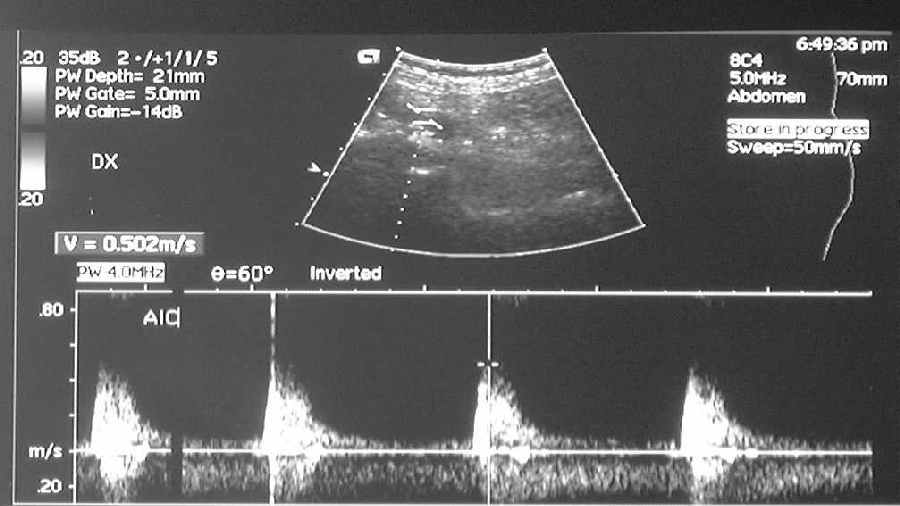
The examination is performed on the pelvic arteries. In each picture the picture on the top shows where the measurement was made. The graphs at the bottom has a velocity scale on the y-axis and a time scale on the x-axis. Notice that the velocity scale is different on the three pictures.
Ultrasound with doppler
Same apparatus is used to make greytone pictures with ultrasound and to measure the bloodstream velocity with doppler. Ultrasound pictures are used to identify arteries and veins, amongs other things, to show stenotic lumen, plaque, sclerotic vessels, thromboses in veins. Doppler examination measures bloodstream velocities in the vessels and presents velocity curves (spectraldoppler). The examination makes it possible to show areas with changed velocities, lacking flow, or change in fow f.ex. vessel with high or low resistance.
Local high velocities in an artery is a typical find in a stenosis. In the legs a velocity increse 2-3 times of a referance area proximal to the stenosis, indicate a significant stenosis with diameter reduction 60-70%.
Doppler examination is less accurate than angiography, but is useful as a primary tool to examine renal artereies and neck arteries, among other things. It is very useful in check-ups after angioplasty or vascular surgery.
Reading
Measurements in the common iliac artery proximal to the stenosis shows a normal graph. The maximum systolic velocity is measured to 0.5 m/sec, and there is no flown in diastole. A measurement in a stenosis in the transition from common iliac artery and external iliac artery (b) shows a maximum systolic velocity of 4.1 m/sec. There is a dramatic increase in the velocity related to the stenotic area. Peripherally to the stenosis the systolic velocity is 0.4 m/sec, while the diastolic velocity is increased.
The finding is typical for a stenosis in the lower extremities. A systolic velocity increase of 2-3 times the referance area proximal to the stenosis, indicates a significant stenosis with a diameter reduction of 60-70%. Ultrasuond with doppler of the entire lower limb is a time consuming examination, and the examination is best suited for a limited area f.ex. restenotsis after previous treatment.
Conclusion
SIGNIFICANT STENOSIS IN THE RIGHT EXTERNAL ILIAC ARTERY.